What happens before the sample hits the cytometer
In most cytometry labs, it's routine to process samples hours, or even days, after collection. But the clock starts ticking the moment blood is drawn. Cells degrade, populations shift, and data quality quietly erodes.
Within 72 hours of the blood draw, fresh blood samples see a:
Experimental design
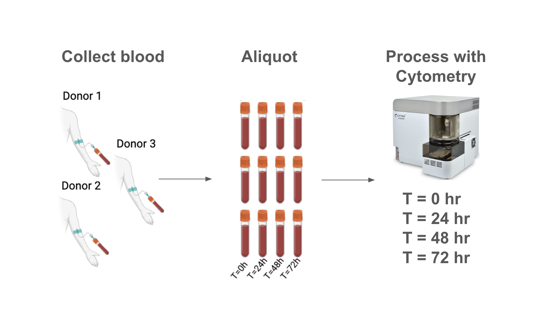
Blood sample degradation measured across 72 hours, in three healthy donors.
Sample collection
Whole blood from three healthy donors was collected in sodium heparin vacutainers. Each specimen was split into eight aliquots: four for live processing and staining, and four for fixation.
Timeline
We analyzed samples at time 0 (immediately after draw), 24, 48, and 72 hours after draw.
Live samples
Samples designated for live processing were diluted in ACK lysis buffer to lyse red blood cells (RBC). Samples were subsequently counted, and stained with a 25-marker antibody cocktail (spectral flow cytometry). After staining, samples were fixed with 1.5% PFA and analyzed.
Analysis equipment
Analysis was performed on a 5 laser Cytek Aurora spectral flow cytometer.
Panel design
Spectral flow cytometry panel design was adapted from the Cytek 25-color Immunoprofiling Assay. The final panel consists of 25 markers with unique spectral signatures and does not contain a viability stain.
By 24 hours, you're already losing nearly 20% of your sample.
Cell loss starts early and does not slow down.
- 15% of cells are lost in 24 hours.
- 33% of cells are lost in 48 hours.
- 48% of cells are lost in 72 hours.
Not all cell populations are lost equally.
Fresh blood samples see variable loss across immune cell populations, making it impossible to predict the patient's true biology at T = 0.
- An 88% drop in monocytes: from ~20% to just 2.7% of nongranulocytes by 72 hours.
- A 48% drop in neutrophils: from ~80% to 40% of total leukocytes over the same period.
CD66b is hit hard in fresh samples, making it difficult to identify neutrophils.
Missed neutrophils lead to distorted immune profiles that inflate T, B, NK, and dendritic cell frequencies.
.gif)
Why does fresh blood sample degradation matter for your team?
Degradation distorts patient immune profiles
When cells die or surface markers decay, immune profiles shift, creating results that reflect technical variation rather than a patient's true biology.
Failed samples waste time and budget
Degraded samples often fail QC, requiring costly redraws, repeat shipments, and delays in data delivery.
Inconsistencies undermine PD insights
Differences in how long samples sit before processing introduce noise, making it harder to detect real pharmacodynamic signals or validate biomarkers.
Want to dig deeper into fresh blood sample degradation?
See this data, compared to the results of samples fixed with TokuKit, in our published whitepaper.